Reading Spine MRI

Neuroradiology series · Part 4 of 4

Anatomic Checkpoints

Two questions to settle before you grade anything — where the cord ends, and whether you are counting the right level.

Companion to Parts 1–3.

1Why this part is last, and why it happens first

Everything in Parts 1 to 3 assumed two things without saying so: that the spinal cord ends where you expect, and that the level you are naming is the level you are looking at. Both assumptions fail often enough to check every time, and both fail silently.

These are quick checks — seconds each, at the start of the study, before any grading. They are last in the series because they only make sense once you know what you are grading, and first in practice because everything downstream depends on them.

Two questions, in this order.
1 · Where does the cord end? If it ends low, you are no longer reading a degenerative study.
2 · Am I counting the right level? If the anatomy is transitional, every level name below it is provisional until you say what you counted from.

2Checkpoint one — where does the cord end?

Step 1 of the lumbar read. Find the tip of the conus on the mid-line sagittal before you look at a single disc.

Where the cord actually ends 944 consecutive adults on MRI. Read the last row first — it is the only one that changes what you do. The two full-width rows split the whole cohort (81.1% + 18.9% = 100%). Indented rows are a subset of the row above them, so the column does not sum to 100. Conus terminates Proportion What it means At or above the L1–L2 interspace 81.1% Four out of five people. This is the ordinary range. of those, ends at the L1 vertebra a subset of the 81.1% above 51.1% The single commonest place — peak density is the lower third of L1. Below the L1–L2 interspace 18.9% Lower than average, and still entirely normal. Do not call this tethering. of those, at or below the L2–L3 interspace a subset of the 18.9% above 0.7% About one person in 140. This is where the finding stops being a variant. The line to remember A conus ending below the mid-body of L2 is outside the normal range at any age. It is not a degenerative finding and it is not incidental — it asks a different question entirely.
Note how wide the normal range is. Nearly one person in five has a conus ending below the L1–L2 interspace and is completely normal. A conus at L2 is not tethering. The threshold that matters is lower and much rarer: at or below the L2–L3 interspace, which was 0.7% of 944 adults.
Why an APP should care, in one sentence. A low conus points at tethered cord — a different disease, a different operation, and a different specialist. It also changes where a spinal anaesthetic can safely be placed, which is the reason this dataset exists in the first place.

3Checkpoint two — are you counting the right level?

This is the least glamorous thing in the whole series and probably the most consequential. Numbering is an assumption, and it is wrong often enough to check.
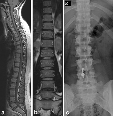
Roughly one spine in nine is not built to the standard plan Whole-spine MRI in 420 consecutive patients. Numbering is an assumption, and it is wrong often enough to matter. 3.3% have a lumbosacral transitional vertebra L5 partly sacralised, or S1 partly lumbarised 7.7% have some other numerical variant extra or missing segments elsewhere in the column The rule that prevents the error Count from a fixed point Start at a landmark you can defend — the last rib-bearing vertebra, or C2 — and count down on one long sagittal. Never count from the gap “The lowest disc” is not a landmark. A sacralised L5 has no mobile disc below it, so the lowest disc may be L4–5. Name your landmark Write it down: “counting from the last rib-bearing vertebra, this is L4–5.” The next reader can then check your count. Flag the variant itself If the anatomy is transitional, say so in the first line. It is not a footnote — it changes what every level below is called. This is a documented “never event”, not a hypothetical A published case: a microdiscectomy carried out one level too high, leaving the original disc untreated, because the patient had ribs at C7 and only eleven thoracic pairs. At the revision, experienced surgeons counting carefully from several landmarks were still uncertain.
Sacralised L5 on whole-spine sagittal MRI, coronal MRI and radiograph
Tins & Balain, Insights Imaging 2016;7:199–203, Fig 1 CC BY 4.0

Sacralised L5 — the lowest disc is not L5–S1

Whole-spine sagittal, panel (a)

The lowest lumbar segment has partly fused to the sacrum. If you count upward from “the lowest mobile disc”, you will name every level one lower than it is.
Notice what panel (a) is: a whole-spine sagittal. This is why counting needs a long sequence or a localiser that reaches a fixed landmark. On a lumbar-only study there may be nothing reliable to count from.
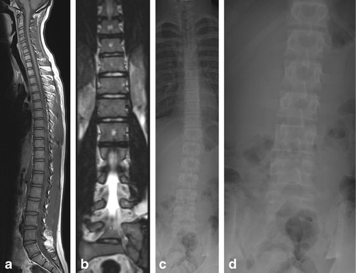
Lumbarised S1 on whole-spine sagittal MRI, coronal MRI and radiographs
Tins & Balain, Insights Imaging 2016;7:199–203, Fig 2 CC BY 4.0

Lumbarised S1 — there is an extra level

Whole-spine sagittal, panel (a)

The opposite error. The top sacral segment has separated and behaves like a sixth lumbar vertebra, giving an extra mobile disc below L5. Count from below here and every level comes out one too high.

The two variants push the count in opposite directions, which is exactly why “count from the bottom” is not a method. Say what you counted from.

4Checkpoint three — is the conus itself the problem?

Parts 1 to 3 were about things pressing on neural tissue from outside. Occasionally the tissue itself is the lesion, and it sits exactly where a routine lumbar study starts.
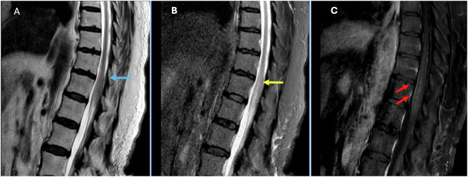
The presentation that should stop you. Saddle anaesthesia, new retention, bilateral leg symptoms — the picture everyone is trained to call cauda equina — can also be produced by a lesion in the conus itself. The symptoms are similar; the cause is not degenerative; the workup is different. If the discs look unimpressive against the symptoms, look hard at the conus before you accept the study as negative.
Sagittal T2, STIR and post-contrast T1 showing an abnormal conus
Sagittal T2, STIR, and post-contrast T1.
Insights Imaging 2025;16:2117, Fig 9 CC BY 4.0

Cauda equina symptoms, conus cause

Sagittal T2 first — then look again with fresh eyes

This man presented with saddle anaesthesia and incontinence. The T2 shows only mild expansion of the conus and the STIR change is described as subtle. It is the contrast study that makes it obvious.
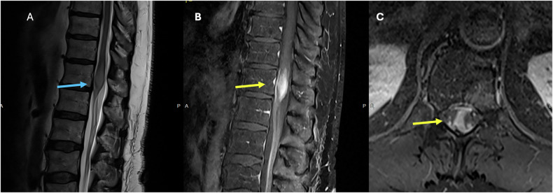
What to take from this. The finding that explained everything was easy to miss on the sequences a routine lumbar study actually contains. You are not expected to make this diagnosis. You are expected to notice that the discs do not explain the patient, and to say so.
Sagittal T2 and post-contrast images of an expanded, enhancing conus
Insights Imaging 2025;16:2117, Fig 4 CC BY 4.0

When it is not subtle

Sagittal T2 · low back pain, numbness and weakness

The same region, and here the cord is frankly expanded with bright signal inside it. Note the presenting complaint: low back pain with leg symptoms — indistinguishable, on the referral, from any degenerative study you will read this week.

Expansion
The cord is fatter than the segment above. Compression makes cord thinner, never fatter. Expansion is always a defer.
Signal inside
Bright T2 within the conus, not around it.
The single rule: a cord that is bigger than it should be is never degenerative.
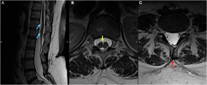
Sagittal and axial T2 showing a syrinx, duplicated dural sac and incomplete posterior fusion
Insights Imaging 2025;16:2117, Fig 12 CC BY 4.0

Congenital findings hide in ordinary studies

A routine lumbar MRI for back pain

A 47-year-old scanned for back pain, with three congenital findings: a small syrinx at T12–L1, a duplicated dural sac split by a midline bony spur, and incomplete fusion of the posterior elements of L5.

None of these is what the scan was ordered for. All of them change the anatomy an operation would encounter, and the split dural sac belongs in the same family as the low conus in checkpoint one.

Why it belongs in this part: these are the findings that are missed precisely because the reader already decided what study they were looking at. The checkpoints exist to interrupt that.

5What you can and cannot call

Call it and act on itDefer it, always
The level at which the conus endsWhy a conus is expanded or enhancing
That the conus is or is not below the L2–L3 interspaceAny intramedullary lesion, of any kind
That the segmentation is transitional, and what you counted fromWhich numbering convention the surgeon should adopt
That the cord looks expanded rather than compressedTumour versus inflammation versus infarct
That the degenerative findings do not explain the symptomsWhat the alternative diagnosis is
The sentence to write. "Counting from the last rib-bearing vertebra, this is a transitional segment with a sacralised L5; levels are named accordingly. Conus terminates at the lower third of L1. No cord expansion or intramedullary signal change." Two sentences, and the next reader knows exactly what you assumed.
Where the series ends. Across four parts the recurring point has not been any grading system — it is that your read exists to raise a question, never to close one. Schizas grades, Pfirrmann grades and Lee grades all describe pictures. The checkpoints in this part describe whether the picture is the one you think you are looking at.

6Where this stops being a teaching point

The counting problem in §3 is not only a reading problem. It is also the reason a whole class of spine AI quietly fails.

A segmentation model trained on ordinary spines learns that the vertebra above the sacrum is L5. Show it a sacralised L5 and it will confidently label the level below as S1 when the surgeon calls it L5 — or show it a lumbarised S1 and every label above shifts by one. The model is not uncertain. It is wrong, and it is wrong in exactly the same direction a hurried human is wrong, so a second reader agreeing with it proves nothing.

That failure mode is invisible unless a dataset contains transitional anatomy and labels it as such. Most public spine datasets do not: transitional cases are uncommon enough that they get excluded during curation, or included and mislabelled by the very convention that the variant breaks.

This is why CTSpinoPelvic1K exists. It is the first CT segmentation dataset for the spine and pelvis built to be LSTV-aware — transitional anatomy is deliberately represented and explicitly annotated rather than quietly dropped. The clinical checkpoint you have just read and the labelling standard the dataset uses are the same idea, one applied by a person and one by a model.
See CTSpinoPelvic1K → OpenSpineConsortium home

The education model that produced this series is described in Schehr A, Kim J, Schwing G, OpenSpineConsortium: an open-source framework for medical student engagement in computational spine imaging research, Cureus 2026;18(7):e112661 (doi:10.7759/cureus.112661).

Sources. Paul JE, et al. Conus medullaris termination: assessing safety of spinal anaesthesia in the L2–L3 interspace. Acta Anaesthesiol Scand 2025;69(3):e14580. — 944 adults on MRI; conus at L1 in 51.1%, at or above the L1–L2 interspace in 81.1%, at or below the L2–L3 interspace in 0.7%.

Tins BJ, Balain B. Incidence of numerical variants and transitional lumbosacral vertebrae on whole-spine MRI. Insights Imaging 2016;7(2):199–203 CC BY 4.0 — 420 consecutive whole-spine MRI studies; transitional lumbosacral vertebra in 3.3%, other numerical variants in 7.7%.

Lindley EM, Botolin S, Burger EL, Patel VV. Unusual spine anatomy contributing to wrong level spine surgery: a case report and recommendations for decreasing the risk of preventable "never events". Patient Saf Surg 2011;5:33 CC BY 2.0 — the wrong-level case described in §3.

Pathology of the conus medullaris and cauda equina: beyond the usual suspects. Insights Imaging 2025;16:2117 CC BY 4.0 — source of the three conus cases in §4.

Every image on this page is openly licensed and clickable.