Model
Weights
YOLO11m-Pose · 1024
Levels
Levels are an assumption about the film, not a model output. Pick a region to
name them; angles that need named endplates appear once you do.
Framing
Confidence
0.50
Trained on 1398 lateral lumbar films (BUU-LSPINE) at fixed hyperparameters with
every augmentation zeroed. Corner order is sup_ant, sup_post, inf_ant,
inf_post and it is handed — mirror the film if anterior and
posterior come out swapped.
On a long or standing film the lumbar spine is a third of the frame, and
a single pass at 640 px finds nothing: measured across 40 re-framed test films,
detections fall from 6.0/6 to 0 once the spine drops below about
two-thirds of the height. Tiling the film into overlapping square windows holds
5.4–5.7 of 6 at every framing down to 0.20, with corner error flat at
0.28–0.33 % of the diagonal. Auto takes the single pass and only pays for
tiles when it comes up short.
Finding the femoral head centre
PI and PT are undefined without the bicoxofemoral axis —
the midpoint of the two femoral head centres (Legaye & Duval-Beaupère,
Eur Spine J 1998). The head is very nearly a sphere, so its projection
is a circle and the landmark is the centre of that circle: trace the
subchondral cortical arc and mark its centre of curvature, not the brightest
shadow — overlap with the acetabulum puts the densest region medial to
the true centre.
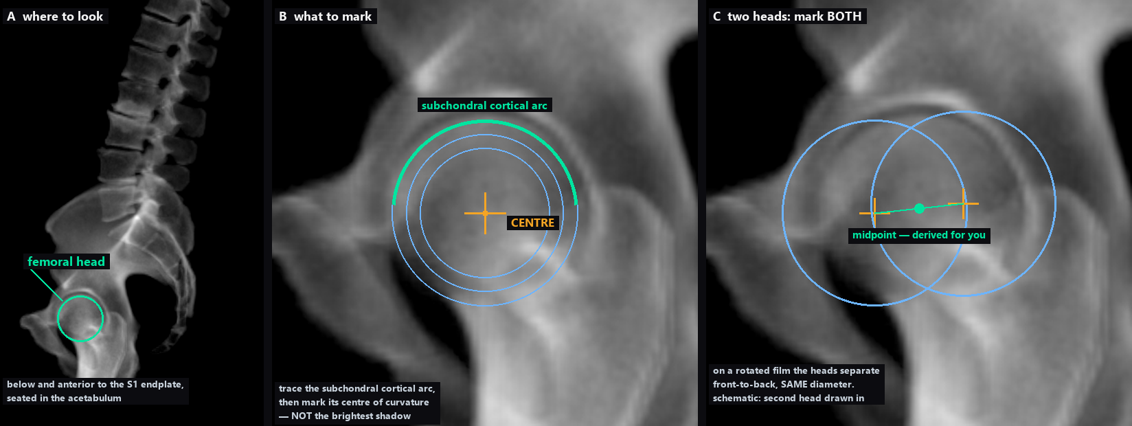 Drawn on the DRR of this case, so the marked centre is ostk's own 3-D sphere
fit projected rather than anyone's opinion. A DRR integrates along the
bicoxofemoral axis, so its two heads superimpose exactly — the second head in
panel C is therefore a schematic, at the same radius and offset front-to-back,
which is how rotation separates them on a real film.
Drawn on the DRR of this case, so the marked centre is ostk's own 3-D sphere
fit projected rather than anyone's opinion. A DRR integrates along the
bicoxofemoral axis, so its two heads superimpose exactly — the second head in
panel C is therefore a schematic, at the same radius and offset front-to-back,
which is how rotation separates them on a real film.
What the film has to contain
SS and LL need only the L1 and S1 endplates, so a coned lumbar
lateral is enough and both are reported. PI and PT additionally
need the femoral head centres; the model has no landmark for them that
survives on a real film, so they are reported as
n/a rather than estimated.
The reference pane is a DRR — the CT from the other tab integrated along
the patient's true bicoxofemoral axis. Its landmarks were fitted in 3-D
and projected, which is why its sacral plate is the true endplate and not the
silhouette of the sacral ala.